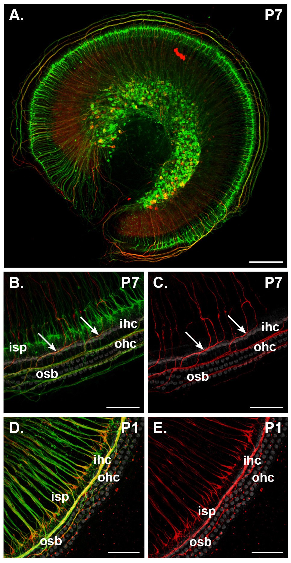
Figure 4

The expression pattern of β-tubulin and peripherin in type I and type II spiral ganglion neurons is maintained in vitro. Maximum intensity projections from confocal z-stacks show immunofluorescence labeling for β-tubulin (green) and peripherin (red) in the mid-apex-mid-turn region of P1 (D,E) and P7 (A-C) mouse cochlea following 48 hour organotypic culture (SGNs and organ of Corti intact). Rhodamine-phalloidin labeling (grey in (B-E)) confirmed the survival of target hair cells. (A,B) Immunolabeling of organotypic cultures of P7 tissue shows the inner spiral plexus (isp), formed by type I fibers, contains β-tubulin protein only (detailed in (B)), whereas the outer spiral bundles (osb) that arise from type II fibers express both β-tubulin and peripherin (arrows in (B,C)). (C) Examination of peripherin immunofluorescence alone confirms that this protein is expressed only in fibers that cross the tunnel of Corti and innervate the outer hair cells (ohc). (D,E) Confocal imaging of organotypic culture of P1 cochlear tissue shows that singularly β-tubulin immunofluorescent fibers beneath the inner hair cells (ihc) are reduced in density compared to the in vivo situation (compare (D) with Figure 1B). A large portion of these fibers co-immunolabel for peripherin (D,E). The outer spiral bundle double immunolabels with β-tubulin and peripherin (D,E). Scale bars: 150 μm (A); 50 μm (C-E).
